神经细胞分化因子1抗体
产品名称: 神经细胞分化因子1抗体
英文名称: Bcl-xL
产品编号: 1336
产品价格: null
产品产地: 上海
品牌商标: 雅吉
更新时间: null
使用范围: WB ELISA IHC-P IHC-F Flow-Cyt IF
- 联系人 :
- 地址 : 上海市闵行区元江路5500号第1幢5658室
- 邮编 :
- 所在区域 : 上海
- 电话 : 158****3937 点击查看
- 传真 : 点击查看
- 邮箱 : yajikit@163.com
| 中文名称 | 神经细胞分化因子1抗体 |
| 别 名 | atonal; Neurod1 protein; basic helix loop helix transcription factor; bHLHa3; class A basic helix loop helix protein 3; Class A basic helix-loop-helix protein 3; MODY 6; MODY6; NDF1_HUMAN; NeuroD1; neurogenic helix loop helix protein NEUROD; Beta cell E box transactivator 2; BETA2; BHF 1; BHF1; NEUROD; Neurogenic differentiation 1; Neurogenic differentiation factor 1; NIDDM; BHLHA3; NEUROD1. |
研究领域肿瘤 心血管 免疫学 染色质和核信号 神经生物学 干细胞 新陈代谢 表观遗传学
抗体来源Rabbit
克隆类型Polyclonal
交叉反应Mouse, Rat, (predicted: Human, Dog, Pig, Cow, )
产品应用WB=1:500-2000 ELISA=1:500-1000 IHC-P=1:100-500 IHC-F=1:100-500 Flow-Cyt=0.2ug/test IF=1:100-500 (石蜡切片需做抗原修复)
not yet tested in other applications.
optimal dilutions/concentrations should be determined by the end user.
分 子 量40kDa
细胞定位细胞核 细胞浆
性 状Liquid
浓 度1mg/ml
免 疫 原KLH conjugated synthetic peptide derived from human NeuroD1:21-120/356
亚 型IgG
纯化方法affinity purified by Protein A
储 存 液0.01M TBS(pH7.4) with 1% BSA, 0.03% Proclin300 and 50% Glycerol.
保存条件Shipped at 4℃. Store at -20 °C for one year. Avoid repeated freeze/thaw cycles.
PubMedPubMed
产品介绍This gene encodes a member of the NeuroD family of basic helix-loop-helix (bHLH) transcription factors. The protein forms heterodimers with other bHLH proteins and activates transcription of genes that contain a specific DNA sequence known as the E-box. It regulates expression of the insulin gene, and mutations in this gene result in type II diabetes mellitus. [provided by RefSeq, Jul 2008]
Function:
Acts as a transcriptional activator: mediatestranscriptional activation by binding to E box-containing promoterconsensus core sequences 5'-CANNTG-3'. Associates with the p300/CBPtranscription coactivator complex to stimulate transcription of thesecretin gene as well as the gene encoding the cyclin-dependentkinase inhibitor CDKN1A. Contributes to the regulation of severalcell differentiation pathways, like those that promote theformation of early retinal ganglion cells, inner ear sensoryneurons, granule cells forming either the cerebellum or the dentategyrus cell layer of the hippocampus, endocrine islet cells of thepancreas and enteroendocrine cells of the small intestine. Togetherwith PAX6 or SIX3, is required for the regulation of amacrine cellfate specification. Also required for dendrite morphogenesis andmaintenance in the cerebellar cortex. Associates with chromatin toenhancer regulatory elements in genes encoding key transcriptionalregulators of neurogenesis (By similarity).
Subunit:
Interacts (via helix-loop-helix motif domain) with EP300(via C-terminus) (By similarity). Heterodimer with TCF3/E47; theheterodimer is inhibited in presence of ID2, but not NR0B2, toE-box element. Efficient DNA-binding requires dimerization withanother bHLH protein. Interacts with RREB1. Interacts with EP300;the interaction is inhibited by NR0B2. Interacts with TCF3; theinteraction is inhibited by ID2.
Subcellular Location:
Cytoplasm. Nucleus. Note=Inpancreatic islet cells, shuttles to the nucleus in response toglucose stimulation. Colocalizes with NR0B2 in thenucleus.
Post-translational modifications:
Phosphorylated. In islet cells, phosphorylated on Ser-274upon glucose stimulation; which may be required for nuclearlocalization. In activated neurons, phosphorylated on Ser-335;which promotes dendritic growth. Phosphorylated by MAPK1;phosphorylation regulates heterodimerization and DNA-bindingactivities. Phosphorylation on Ser-266 and Ser-274 increasestransactivation on the insulin promoter in glucose-stimulatedinsulinoma cells (By similarity).
DISEASE:
Maturity-onset diabetes of the young 6 (MODY6)[MIM:606394]: A form of diabetes that is characterized by anautosomal dominant mode of inheritance, onset in childhood or earlyadulthood (usually before 25 years of age), a primary defect ininsulin secretion and frequent insulin-independence at thebeginning of the disease. Note=The disease is caused by mutationsaffecting the gene represented in this entry.
Similarity:
Contains 1 bHLH (basic helix-loop-helix) domain.
SWISS:
Q13562
Gene ID:
4760
Database links:
Entrez Gene: 4760 Human
Entrez Gene: 18012 Mouse
Entrez Gene: 29458 Rat
Omim: 601724 Human
SwissProt: Q13562 Human
SwissProt: Q60867 Mouse
SwissProt: Q64289 Rat
Unigene: 574626 Human
Unigene: 709709 Human
Unigene: 4636 Mouse
Unigene: 44289 Rat
Important Note:
This product as supplied is intended for research use only, not for use in human, therapeutic or diagnostic applications.
神经生物学相关蛋白(Neurobiology)
| 产品图片 |
 Sample:
Lane 1: Stomach (Mouse) Lysate at 40 ug Lane 2: Spinal cord (Mouse) Lysate at 40 ug Primary: Anti-NeuroD1 (bs-1517R) at 1/1000 dilution Secondary: IRDye800CW Goat Anti-Rabbit IgG at 1/20000 dilution Predicted band size: 49 kD Observed band size: 49 kD  Sample:
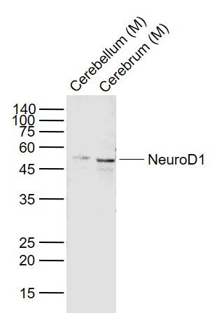
Lane 1: Cerebellum (Mouse) Lysate at 40 ug Lane 2: Cerebrum (Mouse) Lysate at 40 ug Primary: Anti- NeuroD1 (bs-1517R) at 1/2000 dilution Secondary: IRDye800CW Goat Anti-Rabbit IgG at 1/20000 dilution Predicted band size: 49 kD Observed band size: 49 kD Tissue/cell: rat brain tissue; 4% Paraformaldehyde-fixed and paraffin-embedded;
Antigen retrieval: citrate buffer ( 0.01M, pH 6.0 ), Boiling bathing for 15min; Block endogenous peroxidase by 3% Hydrogen peroxide for 30min; Blocking buffer (normal goat serum,C-0005) at 37℃ for 20 min; Incubation: Anti-NeuroD1 Polyclonal Antibody, Unconjugated(bs-1517R) 1:200, overnight at 4°C, followed by conjugation to the secondary antibody(SP-0023) and DAB(C-0010) staining  Blank control: Mouse spleen.
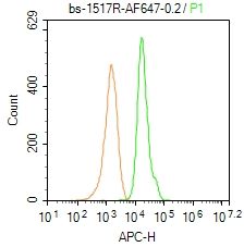
Primary Antibody (green line): Rabbit Anti-Neuro D1/AF647 Conjugated antibody (bs-1517R-AF647) Dilution: 0.2μg /10^6 cells; Isotype Control Antibody (orange line): Rabbit IgG-AF647. Protocol The cells were fixed with 4% PFA (10min at room temperature)and then permeabilized with 90% ice-cold methanol for 20 min at-20℃. The cells were then incubated in 5% BSA to block non-specific protein-protein interactions for 30 min at room temperature. The cells were stained with Primary Antibody for 30 min at room temperature. Acquisition of 20,000 events was performed. |
